Lennart Nilsson, a 91 years-old photographer whose love for science and photography made him very famous. In 1965, LIFE published some amazing pictures on the whole process of how a baby is born. He did it with the help of cystoscope. The first photographs he took were in 1957 which were not clear enough. But in 1965 he finally came up with some beautiful pictures.
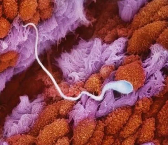
The early stage
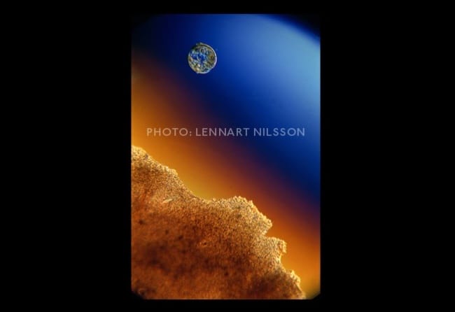
Spermatozoon swam swiftly towards egg via fallopian tube.
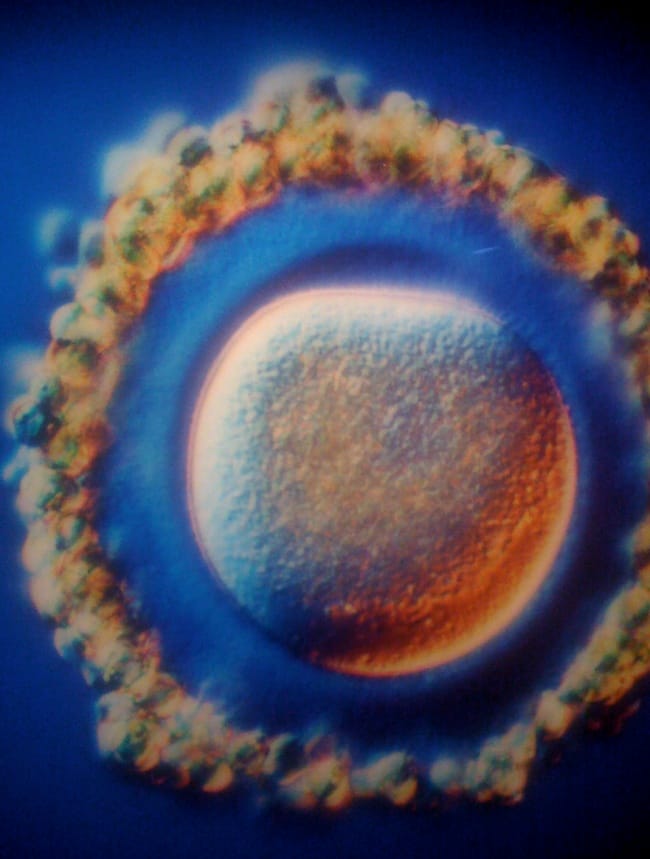
A beautiful female part- an Egg
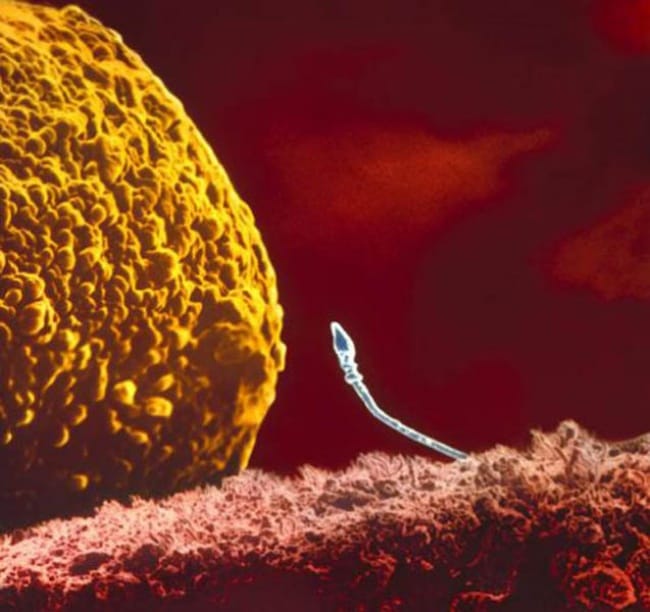
The Attraction
When sperm meets an egg is the crucial moment of a life to begin.
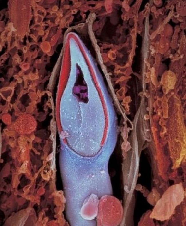
The Entrance
Out of a huge number of sperms one is selected and enters the wall of an egg.
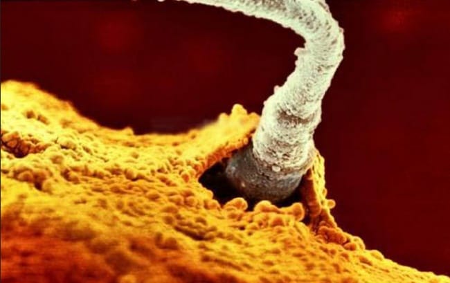
The head of the spermatozoon has all the genetic material.
Baby In Womb
The embryo rolls up from the fallopian tube and shifts to the womb after a week.
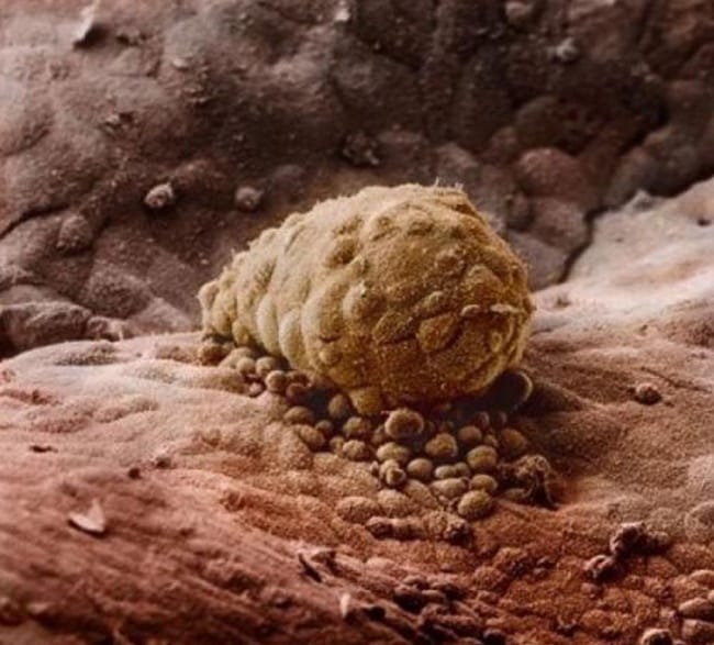
Attachment
1 week later, the embryo gets attached to the wall of the womb
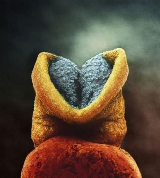
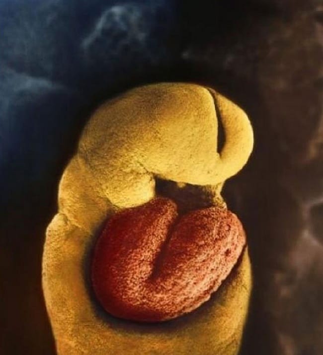
On 22nd day
The grey area turns into brain and this is how 22 days of embryo look like
Beating Heart
On the 18th day, the heart of an embryo is developed.
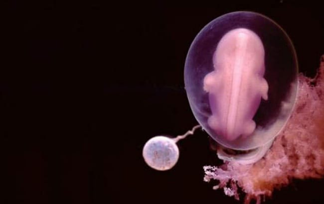
Days Pass By
This is how a cute little fetus looks after 28 days of fertilization.
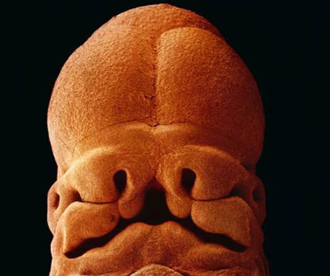
Baby’s Height
After 5 weeks the fetus is about 9 millimeter in size and starts developing the crevices of the face.
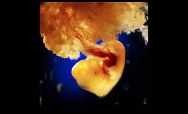
Day 40
When the fetus is 40 days old a placenta is formed.
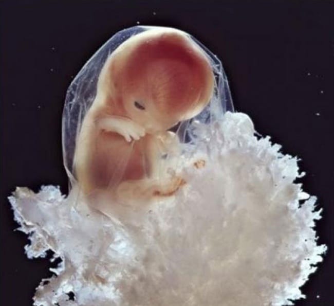
Cute Little Life Inside You
This is an amazing picture of 8 weeks of development.

During 10th Week
On 10th week eyelids are half open and are developed completely within some time.
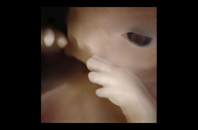
Fetus then starts understanding the inside environment using his/her hands.
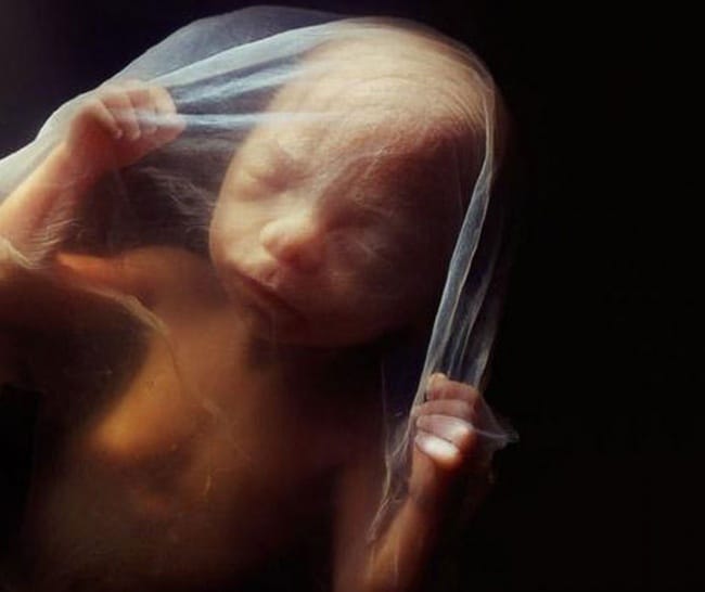
16th Week
At the 16th Week, you can actually see the shape of the full-grown baby.
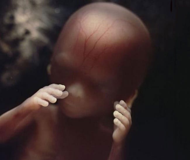
All Developed Nerves
You can observe lines of blood vessels under the skin.
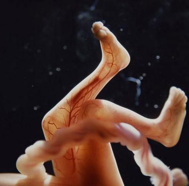
The Listener
On the 18th week, the fetus can start detecting the sound coming from outside.